Arteriovenous Tumor: Dermoscopic Clues
Álvarez-Salafranca, M. (Universidad de Zaragoza) ; Fuentelsaz, V. ; Cebrián García, C.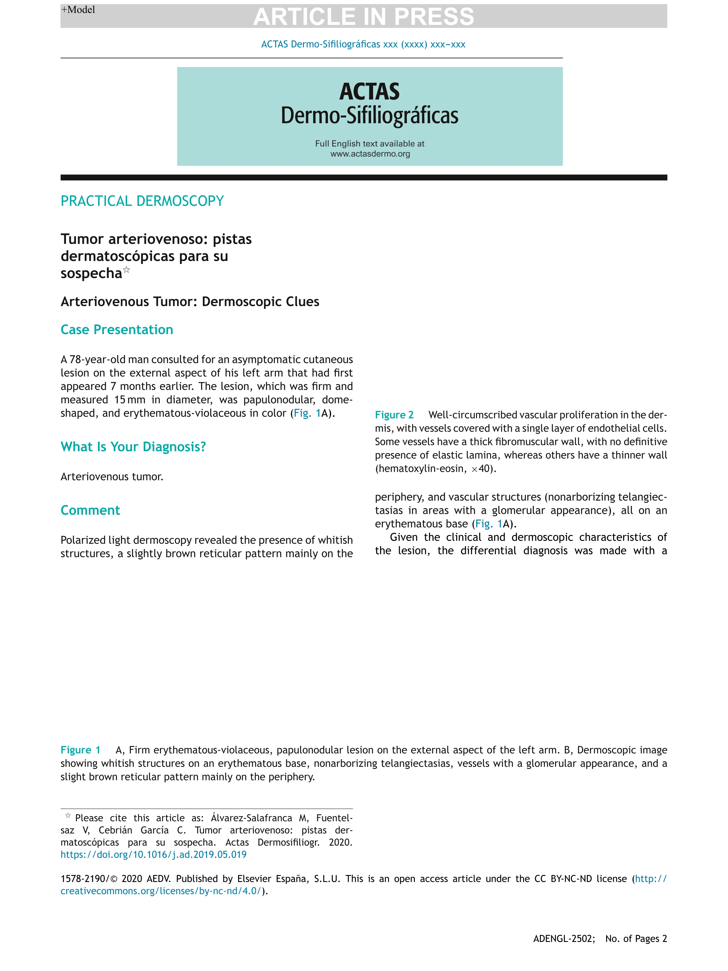
Resumen: Case Presentation
A 78-year-old man consulted for an asymptomatic cutaneous lesion on the external aspect of his left arm that had first appeared 7 months earlier. The lesion, which was firm and measured 15?mm in diameter, was papulonodular, dome-shaped, and erythematous-violaceous in color (Fig. 1A).
Figure 1. A, Firm erythematous-violaceous, papulonodular lesion on the external aspect of the left arm. B, Dermoscopic image showing whitish structures on an erythematous base, nonarborizing telangiectasias, vessels with a glomerular appearance, and a slight brown reticular pattern mainly on the periphery.
What Is Your Diagnosis?
Arteriovenous tumor.
Comment
Polarized light dermoscopy revealed the presence of whitish structures, a slightly brown reticular pattern mainly on the periphery, and vascular structures (nonarborizing telangiectasias in areas with a glomerular appearance), all on an erythematous base (Fig. 1A).
Given the clinical and dermoscopic characteristics of the lesion, the differential diagnosis was made with a vascular lesion, including arteriovenous tumor, aneurysmal dermatofibroma, and hypomelanotic nodular melanoma. ...
Idioma: Inglés
DOI: 10.1016/j.adengl.2021.01.028
Año: 2021
Publicado en: Actas Dermo-Sifiliograficas 112, 4 (2021), 359-360
ISSN: 0001-7310
Factor impacto CITESCORE: 1.2 - Medicine (Q3)
Factor impacto SCIMAGO: 0.279 - Pathology and Forensic Medicine (Q3) - Dermatology (Q3)
Tipo y forma: Article (Published version)
Área (Departamento): Área Dermatología (Dpto. Medicina, Psiqu. y Derm.)
Exportado de SIDERAL (2022-09-08-11:54:57)
Visitas y descargas
A 78-year-old man consulted for an asymptomatic cutaneous lesion on the external aspect of his left arm that had first appeared 7 months earlier. The lesion, which was firm and measured 15?mm in diameter, was papulonodular, dome-shaped, and erythematous-violaceous in color (Fig. 1A).
Figure 1. A, Firm erythematous-violaceous, papulonodular lesion on the external aspect of the left arm. B, Dermoscopic image showing whitish structures on an erythematous base, nonarborizing telangiectasias, vessels with a glomerular appearance, and a slight brown reticular pattern mainly on the periphery.
What Is Your Diagnosis?
Arteriovenous tumor.
Comment
Polarized light dermoscopy revealed the presence of whitish structures, a slightly brown reticular pattern mainly on the periphery, and vascular structures (nonarborizing telangiectasias in areas with a glomerular appearance), all on an erythematous base (Fig. 1A).
Given the clinical and dermoscopic characteristics of the lesion, the differential diagnosis was made with a vascular lesion, including arteriovenous tumor, aneurysmal dermatofibroma, and hypomelanotic nodular melanoma. ...
Idioma: Inglés
DOI: 10.1016/j.adengl.2021.01.028
Año: 2021
Publicado en: Actas Dermo-Sifiliograficas 112, 4 (2021), 359-360
ISSN: 0001-7310
Factor impacto CITESCORE: 1.2 - Medicine (Q3)
Factor impacto SCIMAGO: 0.279 - Pathology and Forensic Medicine (Q3) - Dermatology (Q3)
Tipo y forma: Article (Published version)
Área (Departamento): Área Dermatología (Dpto. Medicina, Psiqu. y Derm.)
Exportado de SIDERAL (2022-09-08-11:54:57)
Permalink:
Visitas y descargas
Este artículo se encuentra en las siguientes colecciones:
articulos > articulos-por-area > dermatologia
Notice créée le 2021-03-09, modifiée le 2022-09-08